Central vs. Peripheral Nervous System: How They Differ and Why It Matters
The nervous system is our body’s lightning‑fast communication network, but it isn’t one monolithic structure. It’s split into two major divisions that each have distinct jobs, protective coverings, and repair capacities. Understanding the key differences between the central and peripheral nervous systems helps demystify everything from concussion symptoms to why a pinprick on the toe feels so sharp.
What Makes the Central Nervous System Unique?
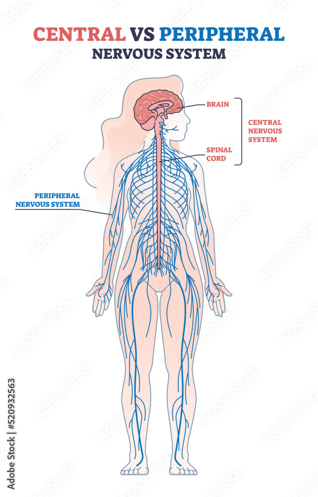
The central nervous system (CNS) consists of the brain and spinal cord. These two structures sit snugly inside the skull and vertebral column, guarded by bone, meninges, and cerebrospinal fluid. Because the CNS processes and integrates almost all incoming sensory data, it’s often described as the “command center.”
- Location and protection: Enclosed by the skull and vertebrae, cushioned by three meninges (dura, arachnoid, pia) and bathed in cerebrospinal fluid.
- Cell types: Dominated by neurons with long axons and a high proportion of glial cells (astrocytes, oligodendrocytes) that support metabolism, maintain the blood‑brain barrier, and insulate axons with myelin.
- Function: Generates thoughts, stores memories, coordinates voluntary movement, and regulates vital autonomic functions like breathing and heart rate.
When the CNS is damaged—say, after a traumatic brain injury—the healing process is notoriously slow. The blood‑brain barrier limits immune cell entry, and many CNS neurons lack robust regenerative capacity.
Peripheral Nervous System: The Body’s Outreach Team
The peripheral nervous system (PNS) branches out from the CNS to every corner of the body. It includes all nerves that run through muscles, skin, and organs, plus the ganglia that house neuronal cell bodies outside the brain and spinal cord.
- Structure: Comprised of cranial nerves (12 pairs that emerge directly from the brain) and spinal nerves (31 pairs that exit the spinal cord). Each spinal nerve splits into a dorsal (sensory) and ventral (motor) root.
- Cell types: Schwann cells are the primary glia, wrapping peripheral axons in myelin and aiding regeneration after injury.
- Function: Relays sensory information from receptors to the CNS (touch, temperature, pain) and carries motor commands back out to skeletal muscles and glands.
Because peripheral nerves sit outside the protective bony encasement, they’re more vulnerable to mechanical stress. On the flip side, they regenerate far more readily—often at a rate of about one millimeter per day—thanks to the supportive environment Schwann cells create.
Key Structural and Functional Contrasts
While both systems use the same basic language of electrical impulses and neurotransmitters, several concrete differences shape how they operate.
| Aspect | Central Nervous System | Peripheral Nervous System |
|---|---|---|
| Primary components | Brain, spinal cord | Cranial & spinal nerves, ganglia |
| Protective covering | Bony skull/vertebrae + meninges + CSF | Connective tissue sheaths (epineurium, perineurium, endoneurium) |
| Dominant glial cells | Astrocytes, oligodendrocytes | Schwann cells |
| Myelin source | Oligodendrocytes (one cell can myelinate multiple axons) | Schwann cells (each cell myelinates a single axon segment) |
| Regenerative capacity | Limited; most neurons do not re‑grow | Relatively robust; axons can often re‑extend |
| Blood‑brain barrier | Present, restricts substance entry | Absent; peripheral capillaries are more permeable |
These contrasts explain why a concussion (CNS injury) can cause lingering cognitive fog, while a nicked fingertip nerve often regains function within weeks.
How the Two Systems Communicate
Signals travel in a two‑step relay: sensory receptors fire peripheral afferent fibers, the impulses sprint to the spinal cord, ascend to the brain for processing, then descend via motor pathways back to the muscles. Think of the PNS as the delivery truck and the CNS as the dispatch center. Disruption at any point—whether a pinched spinal nerve or a stroke in the cortex—can produce very different symptoms.
Clinical Implications of the Differences
Doctors tailor diagnoses and treatments based on whether a problem lies in the CNS or PNS.
- Multiple sclerosis: An autoimmune attack on CNS myelin; peripheral nerves remain largely untouched.
- Carpal tunnel syndrome: Compression of a peripheral nerve (median nerve) causing tingling in the hand, but no brain involvement.
- Spinal cord injury: Often results in permanent loss of sensation and movement below the lesion because CNS neurons rarely regenerate.
- Peripheral neuropathy: Diabetes can damage peripheral nerves, leading to “glove‑and‑stocking” numbness that may improve with glycemic control.
These examples illustrate why imaging, electrophysiology, and symptom patterns are essential tools for pinpointing the affected division.
FAQ
Is the autonomic nervous system part of the CNS or PNS?
The autonomic nervous system is a subdivision of the peripheral nervous system. It controls involuntary functions like heart rate and digestion, though its central coordination hubs reside in the brainstem and hypothalamus.
Can peripheral nerves heal without medical intervention?
Small peripheral nerve injuries often mend on their own thanks to Schwann‑cell‑driven regeneration. Larger gaps may require surgical grafts or nerve transfers to bridge the missing segment.
Why do some drugs affect the brain but not the limbs?
Medications that cross the blood‑brain barrier can act directly on CNS neurons, while others stay in the peripheral circulation and primarily influence peripheral nerves or muscles.
Do CNS and PNS neurons use the same neurotransmitters?
Both divisions employ many of the same chemical messengers—acetylcholine, glutamate, GABA, and norepinephrine, to name a few—though the relative abundance and receptor types can differ markedly.